• We need your support!

    We are currently struggling to cover the operational costs of Xtremepapers, as a result we might have to shut this website down. Please donate if we have helped you and help make a difference in other students' lives!
    Click here to Donate Now (View Announcement)

Physics: Post your doubts here!

Messages
8,477
Reaction score
34,837
Points
698
What is the diff between systematic and random error
Random vs Systematic Error
Random Errors
Random errors in experimental measurements are caused by unknown and unpredictable changes in the experiment. These changes may occur in the measuring instruments or in the environmental conditions.
Examples of causes of random errors are:

  • electronic noise in the circuit of an electrical instrument,
  • irregular changes in the heat loss rate from a solar collector due to changes in the wind.
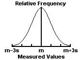
Random errors often have a Gaussian normal distribution (see Fig. 2). In such cases statistical methods may be used to analyze the data. The mean m of a number of measurements of the same quantity is the best estimate of that quantity, and the standard deviation s of the measurements shows the accuracy of the estimate. The standard error of the estimate m is s/sqrt(n), where n is the number of measurements.
ErrorAn2.gif

Fig. 2. The Gaussian normal distribution. m = mean of measurements. s = standard deviation of measurements. 68% of the measurements lie in the interval m - s < x < m + s; 95% lie within m - 2s < x < m + 2s; and 99.7% lie within m - 3s < x < m + 3s.

The precision of a measurement is how close a number of measurements of the same quantity agree with each other. The precision is limited by the random errors. It may usually be determined by repeating the measurements.


Systematic Errors
Systematic errors in experimental observations usually come from the measuring instruments. They may occur because:
  • there is something wrong with the instrument or its data handling system, or
  • because the instrument is wrongly used by the experimenter.
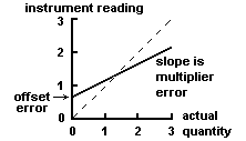
Two types of systematic error can occur with instruments having a linear response:
  1. Offset or zero setting error in which the instrument does not read zero when the quantity to be measured is zero.
  2. Multiplier or scale factor error in which the instrument consistently reads changes in the quantity to be measured greater or less than the actual changes.
These errors are shown in Fig. 1. Systematic errors also occur with non-linear instruments when the calibration of the instrument is not known correctly.
ErrorAn1.gif

Fig. 1. Systematic errors in a linear instrument (full line).
Broken line shows response of an ideal instrument without error.


Examples of systematic errors caused by the wrong use of instruments are:

  • errors in measurements of temperature due to poor thermal contact between the thermometer and the substance whose temperature is to be found,
  • errors in measurements of solar radiation because trees or buildings shade the radiometer.
The accuracy of a measurement is how close the measurement is to the true value of the quantity being measured. The accuracy of measurements is often reduced by systematic errors, which are difficult to detect even for experienced research workers.
 
Last edited:
Messages
62
Reaction score
81
Points
28
Guys Help me

Distinguish between an x-ray image of a body structure and a CT scan?
Don't send me the application booklet, send me just the answer. because the application booklet is not clear


Help me concerning How MRI works and X-ray imaging?


For CT Scan :

Body is divided into sections, each sections is divided into voxels. x ray are directed through the slides at different angels. imag are taken at different angles and are processed and combined together. this is repeated for different slides. image are combined by powerful computers to give a 3d image. the 3d image can be rotated and viewed.

Is it good>
 
Messages
8,477
Reaction score
34,837
Points
698
Guys Help me

Distinguish between an x-ray image of a body structure and a CT scan?
Don't send me the application booklet, send me just the answer. because the application booklet is not clear


Help me concerning How MRI works and X-ray imaging?


For CT Scan :

Body is divided into sections, each sections is divided into voxels. x ray are directed through the slides at different angels. imag are taken at different angles and are processed and combined together. this is repeated for different slides. image are combined by powerful computers to give a 3d image. the 3d image can be rotated and viewed.

Is it good>
X-rays and computed tomography (CT) scans are primarily used to visualize bony structures. CT scans actually use multiple specialized X-rays to view the body area from different angles and then give multiple cross-sectional images of it. The benefits of better visualization offered by CT over X-ray must be weighed against the risks of significantly increased radiation exposure and increased time and costs of the procedure ;

X-rays are a type of radiation, and when they pass through the body, dense objects such as bone block the radiation and appear white on the x-ray film, while less dense tissues appear gray and are difficult to see. X-rays are typically used to diagnose and assess bone degeneration or disease, fractures and dislocations, infections, or tumors.

Organs and tissues within the body contain magnetic properties. MRI, or magnetic resonance imaging, combines a powerful magnet with radio waves (instead of x-rays) and a computer to manipulate these magnetic elements and create highly detailed images of structures in the body. Images are viewed as cross sections or “slices” of the body part being scanned. There is no radiation involved as with x-rays. MRI scans are frequently used to diagnose bone and joint problems.

A computed tomography (CT) scan (also known as CAT scan) is similar to an MRI in the detail and quality of image it produces, yet the CT scan is actually a sophisticated, powerful x-ray that takes 360-degree pictures of internal organs, the spine, and vertebrae. By combining x-rays and a computer, a CT scan, like an MRI, produces cross-sectional views of the body part being scanned. In many cases, a contrast dye is injected into the blood to make the structures more visible. CT scans show the bones of the spine much better than MRI, so they are more useful in diagnosing conditions affecting the vertebrae and other bones of the spine ;

The X-ray and the CT scan both use x-rays.

The X-ray is rather like an ordinary photograph, only the x-rays pass through your body to expose the film behind you instead of the film being in the camera and hit by light waves that bounce off you. That's why the x-ray looks like a film negative: the more x-rays that pass through you, the more exposed is the film and the brighter the image. If you look at an x-ray of a lung with pneumonia, the x-rays pass through the fluid more easily than they do through lung tissue, and so the spaces filled with fluid show up as spidery white lines in the lung.

The CT scan is an x-ray "slice" of your body. It doesn't use film but x-ray sensors that send their signal to a computer as the x-ray transmitter spins around the body to scan a slice of you. Then it moves on to the next slice of you to scan that, sending the x-rays through you to the sensors that send the image to a computer that prints the image, and on it goes.

An MRI is a whole different animal. You are put into a very narrow tube--so narrow that some people have claustrophobia in it even though it is open at the foot and the head--that is filled with a very strong magnetic field. The magnetic field acts on the protons in your body, causing them to "line up" either toward your head or toward your feet, because that' the axis of the magnetic field. Now, the protons line up (actually, they have "spin," and it's more like lining up the axis of their spin) more or less randomly, that is, about half of them line their spins one direction, and the other half line their spins in the other, but not quite. A few more in any area will align their spins in one way, giving that area a slightly more magnetic moment in one direction than in the other. Those are the ones that the machine will detect when a radio frequency pulse resonant to protons is applied to the body. The protons will absorb the energy of that pulse, which the MRI scanner detects. This is called "T1," for "Time 1," the time of energy absorption, and this creates an field that the imager detects. The the protons radiate that absorbed energy at a specific wavelength (which all particles do), and the imager picks up that radiation, which researchers call "T2," for--you guessed it--"Time 2," the time of energy radiation. The MRI can "see" tissues much better than can an x-ray, which can't see soft tissues well at all, except in a few certain circumstances. The MRI uses coils in the tube the patient slides into to selectively apply the magnetic field and the RF pulses to different slices of the body, and it can do so with great precision.

The x-ray technique that can detect bone mineral density is a dual x-ray beam, called DEXA for Dual Energy X-ray Absorbtiometry. 2 x-ray beams of differing energy levels are directed toward the same bone or bones, and the bone's density is calculated from the amount of x-ray energy absorbed from each of the beams.
 
Messages
17
Reaction score
41
Points
13
Hi everyone, I'm kinda having a hard time with testing for homogeneity in some equations.
1. p=rho*g*h......show if this is a homogeneous equation
2.E=mc^2......show if this is a homogeneous equation
Thanks in advance!!!
 
Messages
13
Reaction score
3
Points
13
if the stone has mass m and show that the tension in the cord is given by T=mvw ????????????????????????????????/
 
Messages
12
Reaction score
7
Points
13
Qs- A swimmer who can swim in still water at a speed of 4km h^-1 is swimming in a river. The river flows at a speed of 3km h^-1. Calculate the speed of the swimmer relative to the river bank when she swims:
(a) downstream
(b) upstream


Note: If anyone could upload the answers for Chapters 1 AND 2 for this book

Thanks in advance!
 

Attachments

  • Screen Shot 2014-09-15 at 9.09.59 PM.png
    Screen Shot 2014-09-15 at 9.09.59 PM.png
    303 KB · Views: 4
Top